
Endotracheal Tube (ETT)
The optimal position of ETT can be assessed with CXR. The idea is that you want the tip to be just in between the carina and the vocal cord. Most sources recommend to position the tip of the ETT about 5-7 cm from the carina. Although personally, I try not go higher than 5 cm.
If it too low, there is risk of endobronchial intubation
If it too high, there are risk of the cuff sitting at the level / above the level of vocal cord + risk of accidental extubation.
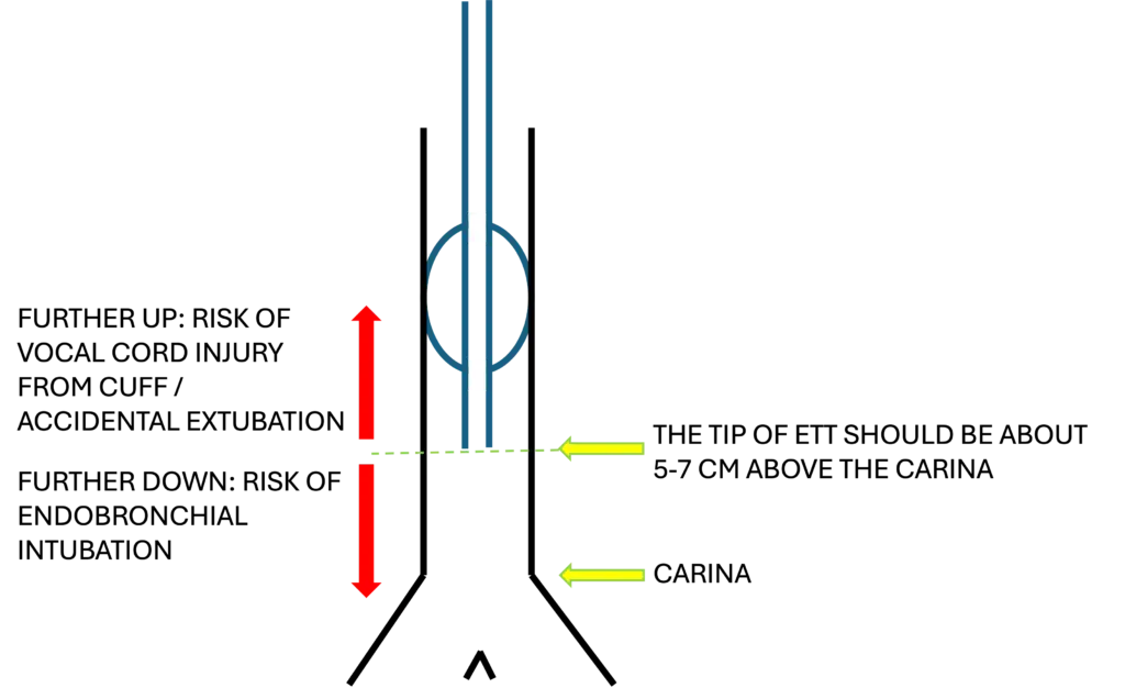
So, do you really need 5 cm space between the tip and the carina? Well, turn out that when the patient moves their neck, the ETT move too. A neck flexion can cause the tip to move as much as 2 cm downwards and a neck extension can cause the tip to move as much as 2 cm upwards. Hence, where the recommendation comes from.
In some cases when you can’t see the carina clearly from the CXR, you can use the vertebral body as a guide. The carina is at approximately at the level of T5 – T7. 5 cm from level of T5 is at the level of T2 which is correlates to the level of sternoclavicular joint.
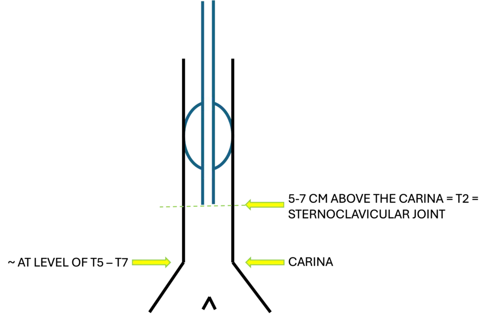
This is a good alternative if you can’t truly see the carina on the CXR. Although I need to remind you that this method is much more prone to error. Patients may have kyphosis or chest wall abnormality so this method would not be reliable.
Some interesting images for more context.
Image 1: The tip of ETT at the ideal level, about 5cm-ish from the carina
Image 2: The tip of the ETT (green line) is positioned slightly higher than the desirable level (above the sternoclavicular joint).
Image 3: The tip of the ETT (green line) is positioned slightly lower than the desirable level (at the right main bronchus).
Key Points:
- The ideal position for the tip is about 5 to 7 cm from carina
- The tip can move with neck flexion / extension
- Consider using vertebral body as guide if you can’t locate the carina on CXR
References
- Bell D, Gaillard F. Evaluation of endotracheal tube position. Radiopaedia. 2025 Jul 27 [cited 2025 Dec 31].
- Edney G, Awal W, Plant L, et al. Vertebral levels (anatomical landmarks). Reference article, Radiopaedia.org (Accessed on 31 Dec 2025) https://radiopaedia.org/articles/57629
- Zaporas I, Thiyagarajan K, Sivasankar R. Postprocedural chest radiograph: Impact on the management in critical care unit. J Mar Med Soc. 2014;16(2):120-124.
- Dundee P, O’Connor M. Chest X-ray – Tubes – ET Tubes – Position. Radiology Masterclass. [updated 2024; cited 2025 Dec 31].
- Ong KC, A’Court GD, Eng P, Ong YY. Ideal endotracheal tube placement by referencing measurements on the tube. Ann Acad Med Singap. 1996 Jul;25(4):550-2. PMID: 8893928. Available from PubMed Central https://pubmed.ncbi.nlm.nih.gov/8893928/
- Goodman LR. Radiographic evaluation of endotracheal tube position. AJR Am J Roentgenol. 1976;127(3):433-434.
Recent Posts
- Right Ventricular Dimension February 27, 2026
- Pneumothorax – CXR February 10, 2026
- The Three Planes / Views January 16, 2026
- The Hounsfield Unit, Window Level & Width January 16, 2026
- THE BLIND MEN & THE ELEPHANT January 14, 2026