

Pneumothorax – CXR What’s normal ? There is not much else to say here. You would expect in a normal CXR with full lungs expansion, you will see the vasculature and bronchial marking spread evenly to the edge of the chest wall bilaterally. What’s not normal ? In CXR with pneumothorax you will see a
Lobar Collapse What’s normal ? For further details on the normal CXR please see specific section here. For quick revision, in the normal CXR where both lungs are fully expanded, (because of the difference in tissue density) you should be able to see all sides border of the lungs, including the normal cardiomediastinal contour. The
Endotracheal Tube (ETT) The optimal position of ETT can be assessed with CXR. The idea is that you want the tip to be just in between the carina and the vocal cord. Most sources recommend to position the tip of the ETT about 5-7 cm from the carina. Although personally, I try not go higher
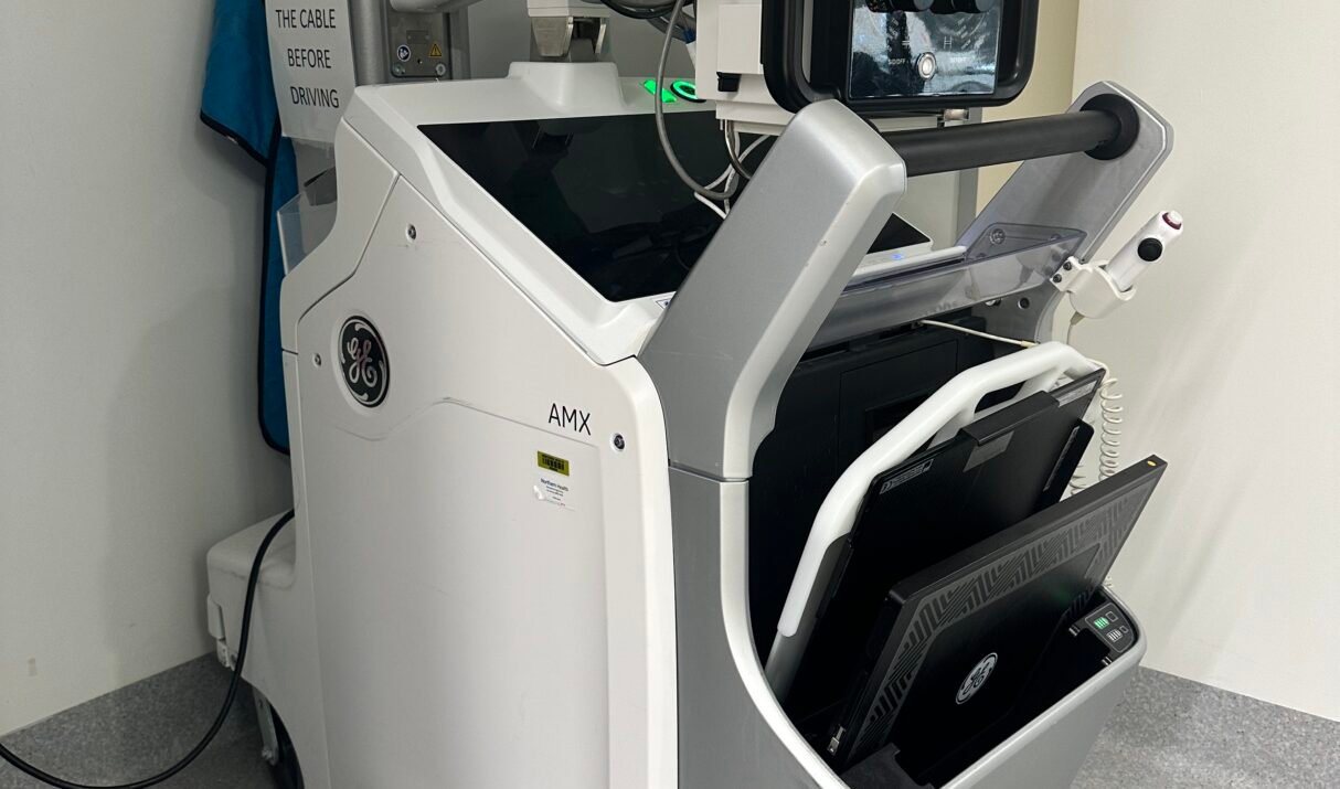
Limitation of Portable CXR Portable Chest X-ray (CXR) in ICU vs. formal CXR in radiology suite What are the differences? Why it matters? Firstly, portable CXR in ICU is done in Anterior-Posterior (AP) position as compared to Posterior-Anterior. If you ask any medical student what’s the difference, the first thing they would say is