
Central Venous Access Device (CVAD)
The optimal position of a central venous access device (CVAD) can be assessed with CXR. The goal is to position the tip at the cavo-atrial junction, which is typically located just inferior to the level of the carina.
If the tip is too high, the risk of thrombus formation increases. This concern is particularly relevant for long-term devices, such as tunnelled vascath. You would also prefer this high-flow access to be placed in larger calibre vessels.
If the catheter tip is positioned too low, below the pericardial reflection, there is a risk of cardiac tamponade due to potential erosion through the vessel wall.
What is the level of risk you might ask? Estimates indicate that among all reported cases of cardiac tamponade, iatrogenic incidents associated with CVC insertion account for a range between 0.0001% and 1.4%. However, there are no reliable resources that indicate a documented risk of cardiac tamponade associated with CVC insertion.
If the tip is placed even lower, within the right atrium, there are increased risks of arrhythmias and damage to the tricuspid valve. The thin wall RA are also more prone to erosion and perforation.
However, the pericardial reflection is not visible on CXR. Therefore, assessment is based on the anatomical location of the carina. Stonelake and Bodenham further divided the area into 3 zones (see image 1).
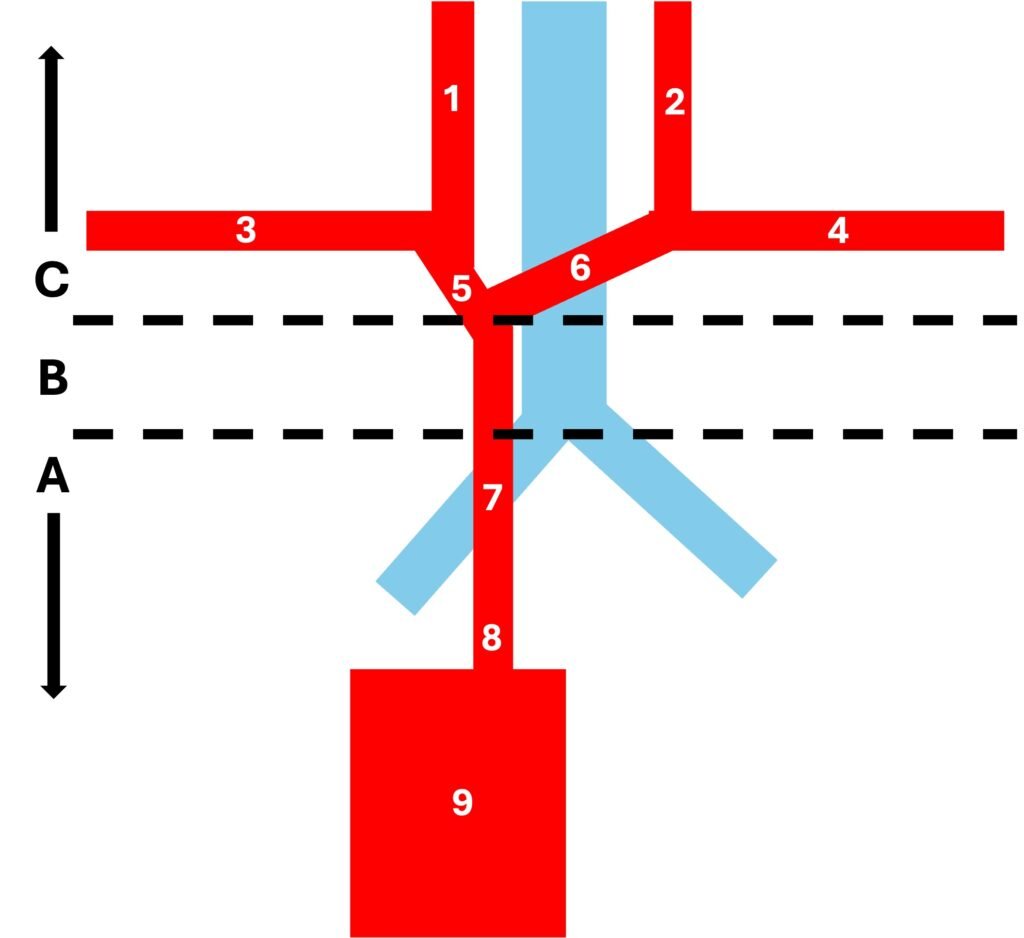
Image 1: 1= Right Internal Jugular Vein, 2 = Left Internal Jugular Vein, 3 = Right Subclavian Vein, 4 = Left Subclavian Vein, 5 = Right Innominate Vein, 6 = Left Innominate Vein, 7 = Superior Vena Cava, 8 = Cavo-atrial Junction, 9 = Right atrium
Zone A: below the carina
Zone B: between the left innominate vein and the carina
Zone C: above the left innominate vein
For a right-side approach, they recommended the tip should in Zone B. For a left-side approach, they recommended the tip should be in Zone A position (to avoid acute abutment to the vein wall).
Below is an example of CVC tip that is in the ideal position on the CXR.
Image 2: Left IJV CVC with the tip at the cavo-atrial junction
Now lets have a look at several examples of positions that may be considered less optimal.
Image 3: Left IJV CVC tip at the left innominate vein (Zone C)
Image 4: Right IJV CVC tip is far too deep in RA
CXR is also useful for identifying malposition, kinking, and coiling—particularly in PICCs—as well as other complications associated with catheter insertion, such as pneumothorax.
Image 5: Right IJV CVC with in the right subclavian vein
Image 6: Right sided PICC coiling back and tip now located in the chest wall. (This is the same patient from Image 3)
Image 7: Left sided PICC with the tip located in the right innominate vein
Key Points:
- Ideal position for the tip is just below the level of the carina
- CXR is also useful to identify malposition, kinking and coiling, and other complications from insertion e.g. pneumothorax
References:
- Bell DJ. CVC position on chest x-ray (summary) Available from: https://radiopaedia.org/articles/cvc-position-on-chest-x-ray-summary
- Radiology Masterclass. Chest X-ray – Tubes – CV Catheters – Position. Radiology Masterclass; Available from: https://www.radiologymasterclass.co.uk/tutorials/chest/chest_tubes/chest_xray_central_line_anatomy
- Venkatesan T, Sen N, Korula AM, Raj JP, S S, Paul TV, et al. Role of chest X-ray in citing central venous catheter tip. J Anaesthesiol Clin Pharmacol;31(3):393-7. Available from: https://pmc.ncbi.nlm.nih.gov/articles/PMC3788245/
- Singh A, Bajpai M. Pictorial essay: central venous catheters on chest radiographs. BJMP;4(4):a448. Available from: https://www.bjmp.org/content/pictorial-essay-central-venous-catheters-chest-radiographs
- Stonelake PA, Bodenham AR. The carina as a radiological landmark for central venous catheter tip position. Br J Anaesth. 2006 Mar;96(3):335-40. Available from: https://www.sciencedirect.com/science/article/pii/S0007091217352054
- Dulay M, Sahi IG, Erbel T, et al. Topographic analysis and evaluation of anatomical landmarks for central venous catheter placement. Br J Anaesth. 2014;112(2):271-80. Available from: https://www.sciencedirect.com/science/article/pii/S0007091217319153
- Schuster M, Nave H, Piepenbrock S, Pabst R, Panning B. The carina as a landmark in central venous catheter placement. Br J Anaesth. 2000 Aug;85(2):192-4. PMID: 10992822. Available from: https://pubmed.ncbi.nlm.nih.gov/10992822/
- Vedran Premuzic, Drazen Perkov, Ranko Smiljanic, Bruna Brunetta Gavranic, Bojan Jelakovic; The Different Impacts on the Long-Term Survival of Tunneled Internal Jugular Hemodialysis Catheters Based on Tip Position and Laterality. Blood Purif11 April 2017; 43 (4): 315–320. https://doi.org/10.1159/000454670
- Kim EH, Moolchandani P, Patel S. Digging Deeper to Diagnose: Cardiac Tamponade Following Tunneled Dialysis Catheter Placement. Cureus. 2024 Sep 27;16(9):e70317. doi: 10.7759/cureus.70317. PMID: 39463659; PMCID: PMC11512760.
- Messina Alvarez AA, Bilal MA, Manasrah N, Chaudhary A. Iatrogenic Cardiac Tamponade Secondary to Central Venous Catheter Placement: A Literature Review. Cureus. 2023 Apr 17;15(4):e37695. doi: 10.7759/cureus.37695. PMID: 37206520; PMCID: PMC10191201.
- Azevedo AC, Flor de Lima I, Brito V, Centeno MJ, Fernandes A. Tamponamento cardíaco: uma complicação rara da cateterização venosa central – relato de um caso clínico [Cardiac tamponade: a rare complication of central venous catheter – a clinical case report]. Braz J Anesthesiol. 2018 Jan-Feb;68(1):104-108. doi: 10.1016/j.bjan.2016.02.003. Epub 2016 Mar 22. PMID: 27016189; PMCID: PMC9391675. Available at:https://pmc.ncbi.nlm.nih.gov/articles/PMC9391675/
Recent Posts
- Piezo 101: Squeezing Electricity Since 1880 April 16, 2026
- Central Venous Access Device (CVAD) April 11, 2026
- Pneumonia – CXR March 28, 2026
- Right Ventricular Systolic Function March 7, 2026
- Right Ventricular Dimension February 27, 2026